THIS ARTICLE IS MORE THAN FIVE YEARS OLD
This article is more than five years old. Autism research — and science in general — is constantly evolving, so older articles may contain information or theories that have been reevaluated since their original publication date.
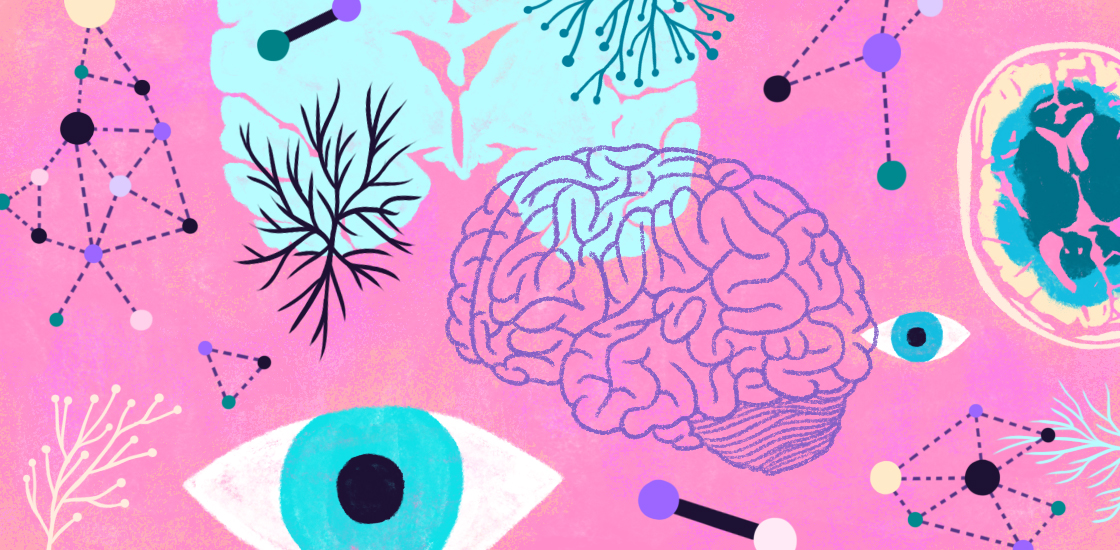
If one were to summarize the state of brain research on autism in a sentence, it might be: “We have many findings, but little understanding.” Neuroimaging research on autism has been enormously productive in the past decades, including tens of thousands of results. But few of the studies have been replicated. We have therefore not been able to piece together a coherent picture of crucial brain features underlying autism.
One problem is heterogeneity: Children diagnosed with autism fulfill the behavioral criteria in the latest version of the “Diagnostic and Statistical Manual of Mental Disorders,” the DSM-5, but do not all have the same biological condition. ‘Autism’ probably includes many different neurobiological conditions that differ in their causes.
A foremost clinical goal in autism research is, therefore, not so much to find the cause and the treatment for the condition, but to find out whether there are biologically distinct types of autism, each potentially treatable in a different way. Consequently, we need to understand whether neuroscientific results tell us something important about ‘autisms’ and their differences — as opposed to findings that only reflect differences in the way we conduct our studies. Research on brain connectivity may serve as an example.
Critical choices:
There is growing agreement that autism is not located in a specific part of the brain, but rather relates to the way different brain regions ‘talk’ to each other. A prominent technique in the study of brain-network communication is functional-connectivity magnetic resonance imaging (fcMRI), which tests whether activity changes are coordinated across different parts of the brain.
About 10 years ago, there appeared to be firm evidence that the brain in autism is underconnected — less coordinated than the neurotypical brain. But then, more and more published studies appeared to show the exact opposite.
As it turns out, different studies used slightly different methods. One investigation in 2014 demonstrated that the same data from the same children could show either underconnectivity or overconnectivity in the brains of children with autism, depending on how the data were analyzed1.
This inconsistency does not mean that fcMRI is not a useful technique. In fact, we can learn much from it, but only if we carefully consider how our methodological choices give us insight into brain function from different angles.
Going local:
Most fcMRI studies in autism to date have examined the connectivity between distant parts of the brain. Much less is known about local connectivity — how neighboring brain sites ‘talk’ to each other — which is equally important.
Neighboring parts of the brain usually play similar roles. For example, regions at the very back of the brain have visual functions, and neighboring regions immediately in front of them are also visual. But these areas specialize: The ones farther back respond to simple shapes such as edges, and the ones a bit farther in front recognize complex forms such as faces.
Researchers can use local-connectivity fMRI to examine both coordination between neighboring brain sites and their distinct roles.
In autism research, a frequent refrain has been that the brain is locally overconnected but underconnected at greater distances. For example, the idea goes, the frontal lobes talk a lot amongst themselves, but too little with far-off parts. Such ideas may be repeated by one review paper after another until they are considered firmly established. But in reality, the question has not been thoroughly examined.
We tested the idea in a study published this year that included data from more than 300 children and adolescents (147 with autism), and found rather different patterns2. First, several brain regions unexpectedly showed local underconnectivity. These were mostly at the midline of the brain in the cingulate cortex, which is important for attention, memory and emotions. In other words, neuronal activity seems to be less coordinated in people with autism, with some neighboring sites talking too little to each other.
Second, there was some local overconnectivity, as expected, but it occurred only in visual regions. This finding could be related to the special status of vision in some people with autism, leading to superior abilities in tasks such as finding a single object in a large array of other objects3.
Open or closed:
Our third main finding concerns the way we commonly run resting-state fMRI scans. In these studies, participants have no task, but are simply asked to relax and lie still for five or more minutes of scanning. However, in previous autism fcMRI studies, one crucial factor has been overlooked: Participants may either keep their eyes open — often to focus on a cross on a screen — or close their eyes.
We found that eye status (open or closed) had a dramatic effect on local connectivity. Visual regions at the back of the brain had much greater local connectivity when participants kept their eyes open, and the group differences in these regions described earlier disappeared.
This relates to general uncertainties about the brain ‘at rest.’ Resting-state fMRI has great advantages, especially because it allows the study of children who may not be able to perform tasks during scans. However, we do not know what exactly is happening in the minds of children when they rest. Specifically, when participants close their eyes, it is possible that they doze off — which is a problem because sleep onset greatly changes activity patterns across the brain4.
Mixing data from eyes-open and eyes-closed scans may therefore give us misleading results of what appear to be differences between people with autism and control participants.
Methods matter:
Much of the above may appear to be scientific fine print that has little to do with the treatment needs of children with autism. But as long as we don’t understand what exact brain patterns are responsible for the problems these children face in their everyday lives, improving our ability to study the brain and to interpret findings from research studies remains crucial.
In other words, if we can’t tell what findings reflect true differences and what findings result from methodological issues, we will not be able to assemble an adequate and coherent picture of brain differences in autism.
This goal will be hard to achieve, because there are probably many ‘autisms,’ each associated with different brain patterns. Continuous fine-tuning of basic science methods, as in brain imaging, and improved understanding of what may derail analyses remain crucial in this endeavor.
Ralph-Axel Müller is professor of psychology at San Diego State University in California.
By joining the discussion, you agree to our privacy policy.