THIS ARTICLE IS MORE THAN FIVE YEARS OLD
This article is more than five years old. Autism research — and science in general — is constantly evolving, so older articles may contain information or theories that have been reevaluated since their original publication date.

The man known for having the ‘most studied brain’ would like to share his title: Psychologist Russell Poldrack says scanning the brain of a single individual over months to years may reveal daily fluctuations in activity that are relevant to conditions such as autism.
Beginning in 2012, Poldrack, then director of the Imaging Research Center at the University of Texas at Austin, scanned his own brain using functional magnetic resonance imaging (fMRI) for 10 minutes, two to three times each week. He kept up the routine for 18 months and, by the end, found distinct differences in his brain activity from one day to the next1.
This high-tech navel-gazing ultimately led to a detailed map of the ever-changing relationships between brain regions — at least in Poldrack’s head. Two brain regions that communicate one day might not be on speaking terms the next.
His idea inspired many others. One group, which includes some investigators from Poldrack’s study, has created a public database of brain maps from 10 typical adults, each of whom were scanned using fMRI for at least five hours.
We asked Poldrack, now professor of psychology at Stanford University, about his findings and how longitudinal brain maps of individuals can advance research on conditions such as autism.
Spectrum: Why did you decide to scan your own brain?
Russell Poldrack: I became interested in how basic cognitive functions relate to psychiatric disorders, and how these functions might span different conditions.
I actually don’t know the degree to which this is the case in autism, but certainly in conditions such as depression, psychosis and bipolar disorder, you see this incredible variability over time. A person can go from being fully functional to fully disabled over the course of a couple of weeks. And it occurred to me that we didn’t have any way to understand what was going on over that period on the basis of cognitive neuroscience.
My intuition was, if we want to understand fluctuations over time in people with a condition, the first thing we need to do is understand them in typical people.
S: Had anyone looked at day-to-day changes in brain activity before your study?
RP: We knew a little bit about very long-term changes across child and adolescent development and aging. And we knew a little bit about changes in brain activity over seconds and minutes. But basically, there had been no work that examined changes over days to weeks to months in individuals. Most researchers look at people once and assume they have a valid picture of those individuals.
Think about that in the context of neuropsychiatric conditions. Take somebody with a diagnosis of schizophrenia. The idea that you will capture their condition with a snapshot of brain activity on the day they sit in the scanner is ludicrous, when you think about it.
S: What are the trade–offs of studying one person over time compared with studying many people at a single point in time?
RP: If we want to understand differences between people — which we often do — then there’s no way around doing large aggregate studies of many people. But the limitation of those studies is that we’re not respecting the fact that individuals vary from day to day. And we’re not getting enough data on individuals to really understand what is going on in their brains.
I think there’s certainly a trade-off between depth and breadth. I think we have to push to do both kinds of studies if we want to answer all of questions we’re interested in.
S: What was your most intriguing finding?
RP: Aggregate data on large groups of people has led other researchers to identify large-scale networks that function in the brain’s resting state — that is, when a person is not actively performing a task.
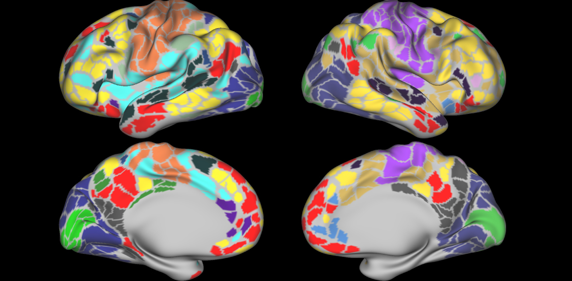
Group studies have generally found that voxels — a unit of a brain scan — throughout the ventromedial prefrontal cortex show connectivity with voxels in the posterior cingulate and the lateral temporo-parietal areas. Together, these regions make up the ‘default mode network,’ which is particularly engaged when the brain is at rest.
In our study, we had about 840 minutes of resting-state data from my brain. What we saw is that my global organization looks roughly like what the group studies have shown. There’s a default mode network in roughly the right place, but there is all this heterogeneity. For example, within the middle of the default mode network there was a little spot of brain connected to different networks that the default mode network does not traditionally connect to, like the salience network.
That was totally unexpected based on the group data. It could be that it was just my weird brain or that’s how brains are. But a study published in August from Nico Dosenbach’s group at Washington University in St. Louis led to similar findings in other individuals1.
S: Are individual brain scan studies useful for understanding autism?
RP: Assuming there’s variability in day-to-day behavior in autism just as there is for other conditions, it will give you a direct way of relating brain systems to that variability. If we do this across individuals, we can see if a person who has severe symptoms of a certain type shows more activity or less activity in a particular brain area.
Scanning a person’s brain repeatedly over a period of time can help us determine if a pattern of brain activity characterizes that person, or is there one day and gone the next. If we can relate that activity directly to an individual over time, then we have a powerful lever for understanding what’s driving the daily changes in brain activity.
I would say researchers should also just start thinking about whether the amount of data that they’re collecting takes into account the variability in the people they’re collecting the data from.
S: Scanning someone’s brain every day sounds arduous. What are the challenges?
RP: For me it wasn’t terrible because I was going to my place of work every day anyway. But the approach is likely to be challenging in many cases. If it’s a person who has to go across town and go park at an imaging center and wait in a waiting room to get an MRI scan regularly, that would be more difficult.
And if it’s a child with autism, her parents are going to be more likely to bail on the scan on days she is acting out. And a person who has depression is less likely to show up for a scan on a day when he or she is deeply depressed. These kinds of selection effects could seriously bias the data.
S: What’s next for your lab?
RP: We have submitted a grant proposal for a detailed fMRI study of 55 typical individuals. We would bring these individuals in multiple times over months to do a large number of tasks that relate to executive function. This is a set of important cognitive skills that includes short-term memory and self-control. What we want to do is obtain an estimate of how each individual’s brain responds to a large set of executive-function tasks. Scanning the participants repeatedly will allow us to obtain these estimates with much higher precision than if we simply scanned them once.
By joining the discussion, you agree to our privacy policy.