THIS ARTICLE IS MORE THAN FIVE YEARS OLD
This article is more than five years old. Autism research — and science in general — is constantly evolving, so older articles may contain information or theories that have been reevaluated since their original publication date.
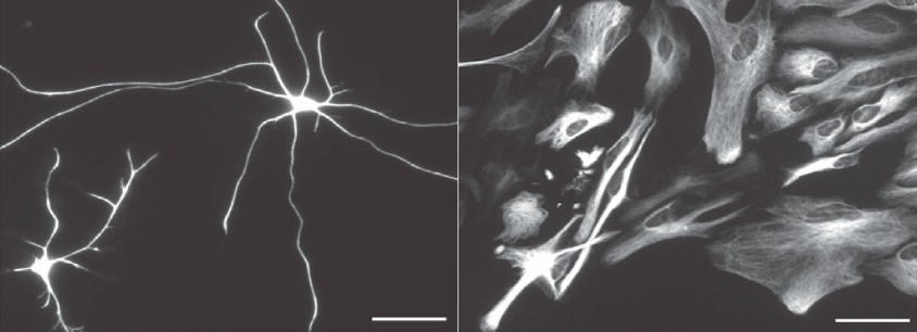
A new method allows researchers to isolate and culture cells known as astrocytes from human brains. The method, detailed 6 January in Neuron, could provide clues about whether anomalies in astrocytes contribute to autism1.
Astrocytes are star-shaped cells that provide nutrients to neurons, among other functions. They make up roughly 40 percent of cells in the brain. Studies in mice and rats suggest the cells help form and prune the junctions between neurons, called synapses. Scientists have linked problems with synaptic pruning to autism.
Researchers were previously unable to extract mature astrocytes from human brain tissue and maintain them in culture. Instead, they relied on immature precursor cells, some of which differentiate into other cell types. They cultured these immature cells in serum, the portion of blood that contains growth factors, nutrients and other molecules to support cell growth. But serum causes astrocytes to flatten and lose their star shape.
The new method allows researchers to isolate astrocytes from brain tissue removed during surgery for conditions such as epilepsy or cancer.
In the technique, the researchers first separate the cells by bathing brain tissue in fluid containing a papaya enzyme that gently breaks down proteins connecting brain cells. Next, they isolate astrocytes from other cell types with a method called immunopanning, which involves using a series of antibody-coated culture dishes to sequentially siphon off immune cells called microglia, then other neural support cells, blood vessel-forming cells and neurons.
The final dish contains antibodies that stick to a protein on the outer membrane of astrocytes. Cells that do not stick are washed away. The cells grow in a medium containing a growth factor and nutrients that do not distort the cells’ shape.
Star players:
The researchers used the method to isolate astrocytes from 12 people who had undergone brain surgery for epilepsy. The cells came from samples of healthy tissue that had been removed as the surgeons cut their way to the epileptic lesions.
The researchers cultured the mature astrocytes with rat neurons. These neurons formed three to four times as many synapses as ones grown without astrocytes.
The researchers also incubated some astrocytes with isolated nerve endings that make up synapses. The astrocytes engulfed these neuronal morsels, suggesting that they’re also involved in synaptic pruning. These findings are in keeping with a role for astrocytes in both building neuronal circuits and fine-tuning them.
Other experiments revealed that human astrocytes express many genes at much higher levels than mouse astrocytes do. Looking at these genes may help scientists understand where mouse astrocytes fall short as models for human disease, the researchers say.
An abbreviated version of the method can isolate astrocyte progenitor cells from fetuses, which contain a limited number of brain cell types. The researchers also aim to use the method to isolate and culture astrocytes from three-dimensional ‘mini-brains’ derived from stem cells.
The method may help scientists better understand the role of astrocytes in the developing brain — possibly yielding clues about how astrocytes contribute to conditions such as autism.
Gene expression data on the isolated human astrocytes and other brain cells is available in the researchers’ online database, Brain RNA-Seq.
By joining the discussion, you agree to our privacy policy.