THIS ARTICLE IS MORE THAN FIVE YEARS OLD
This article is more than five years old. Autism research — and science in general — is constantly evolving, so older articles may contain information or theories that have been reevaluated since their original publication date.
A new technique acts as a genetic geolocator, pinpointing the precise location of active genes in the brain1. The method could reveal atypical patterns of gene expression in brain tissue from individuals with autism.
Existing methods can identify active genes in tissue by sequencing messenger RNA (mRNA) — the molecular intermediate between DNA and protein. But these techniques typically require blending tissue samples into a slurry, which obscures the original locations of the genes.
With the new method, described 1 July in Science, researchers sidestep this problem by identifying each mRNA while it is still embedded in tissue. They start with glass slides coated with millions of molecular probes, each composed of a unique ‘barcode’ sequence that corresponds to a specific location on the slide and a short sequence that sticks to mRNA.
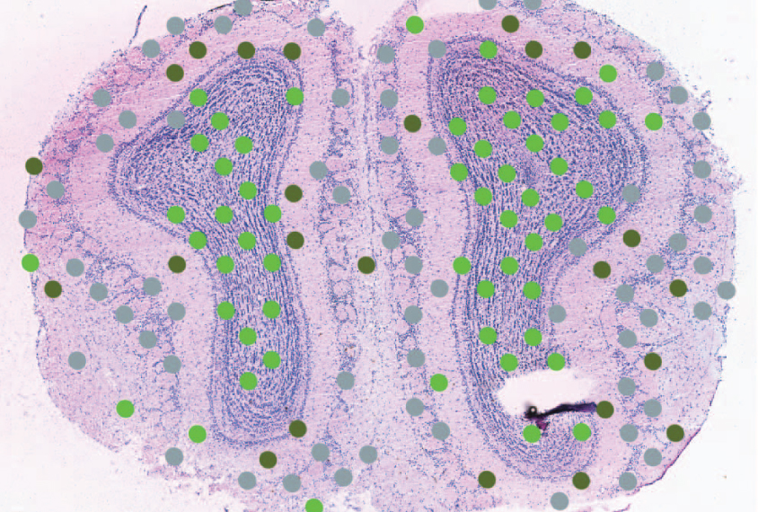
They then mount thin slices of brain tissue onto the slides. They stain the tissue with a standard dye that highlights the shape of cells and reveals patterns in the tissue, such as layers of neurons. The researchers take snapshots of the stained tissue through a microscope to provide a visual template on which they can map gene expression data.
Next, they wash the slides with enzymes that punch holes in cell membranes, causing mRNA to leak out of the tissue onto the slide. The probes snag the corresponding mRNA strands, which researchers collect and sequence using standard techniques. The results reveal which genes were active in the tissue.
The sequences also include barcode information, which points to where on the slides the mRNA was captured. The researchers then match that location with snapshots of the intact tissue.
Using the method, the researchers pinned the activity of 170 genes to particular layers of the mouse olfactory bulb. They also mapped gene expression in thin sections of tissue from a breast cancer biopsy. They found clusters of active genes that may reflect different lineages of the original tumor.
Scientists could use spatial information about gene expression to uncover region-specific changes associated with conditions such as autism. In this study, the researchers detected more than 14,000 active genes in different experiments. Scientists can create slides that recognize thousands of select mRNAs using a kit.
By joining the discussion, you agree to our privacy policy.