THIS ARTICLE IS MORE THAN FIVE YEARS OLD
This article is more than five years old. Autism research — and science in general — is constantly evolving, so older articles may contain information or theories that have been reevaluated since their original publication date.
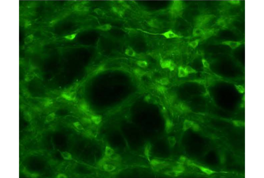
A new method uses luminescent proteins to control neurons in the brains of freely moving rats. The proteins, called luminopsins, light up the brain from within, allowing researchers to dampen the activity of light-sensitive neurons without having to insert invasive probes1.
The method, described 24 September in Scientific Reports, is a variation on the popular optogenetics method, in which researchers engineer rodents to express light-sensitive proteins, called opsins, in some neurons. Surgically inserted fiber-optic cables then transmit light to the neurons, which either ramp up or temper their activity.
Researchers have used optogenetics to study the role of specific neurons in autism-linked behaviors. But the surgery to implant the probes can damage neurons or lead to infection, and the fiber-optic cables can interfere with social behaviors.
In the new study, researchers created a luminopsin protein by combining an opsin with luciferase, the enzyme that gives bioluminescent organisms their glow. They then engineered rats to express the glowing proteins in neurons in the basal ganglia, a brain region that controls movement. They targeted the basal ganglia on one side of each animal’s brain.
The researchers found that when they inject the rats with a chemical that luciferase needs to glow, the luciferase lights up. The attached opsins then pump chloride ions into the neurons. Negative charge builds up in the neurons, preventing them from firing. (In unpublished work, the researchers have designed luminopsins that stimulate neurons using excitatory opsins.)
Injecting these rats with amphetamine, a stimulant, causes the animals to run in circles. The amphetamine is likely to increase neuron firing on one side of the brain while the luminopsins block firing on the other side.
The technique may allow researchers to control neurons in multiple brain regions simply by expressing luminopsins in the neurons, without having to implant multiple fiber-optic cables. For example, luminopsins might be able to inhibit neurons that spark seizures in people with epilepsy, the researchers say.
However, because the chemical trigger for luciferase remains in the body for more than an hour, the technique does not allow researchers to turn neurons off and on rapidly — an advantage of using traditional optogenetics.
By joining the discussion, you agree to our privacy policy.