THIS ARTICLE IS MORE THAN FIVE YEARS OLD
This article is more than five years old. Autism research — and science in general — is constantly evolving, so older articles may contain information or theories that have been reevaluated since their original publication date.
A curved glass replacement for the top of a mouse’s skull lets researchers spy on the activity of more than 1 million neurons1. The device, dubbed ‘the crystal skull,’ offers an unprecedented view of brain activity in living mice.
Scientists typically watch the brain in action by engineering mice with neurons that glow when they fire. They swap a small piece of skull for a glass window, which allows them to see the flickering of neuron chatter through a microscope.
But these windows are usually flat, and unless they are small, press against the brain’s curved surface and damage tissue. Researchers can sidestep this problem by thinning the skull until it is transparent. But even thinned bone can obscure the glow from neurons below.
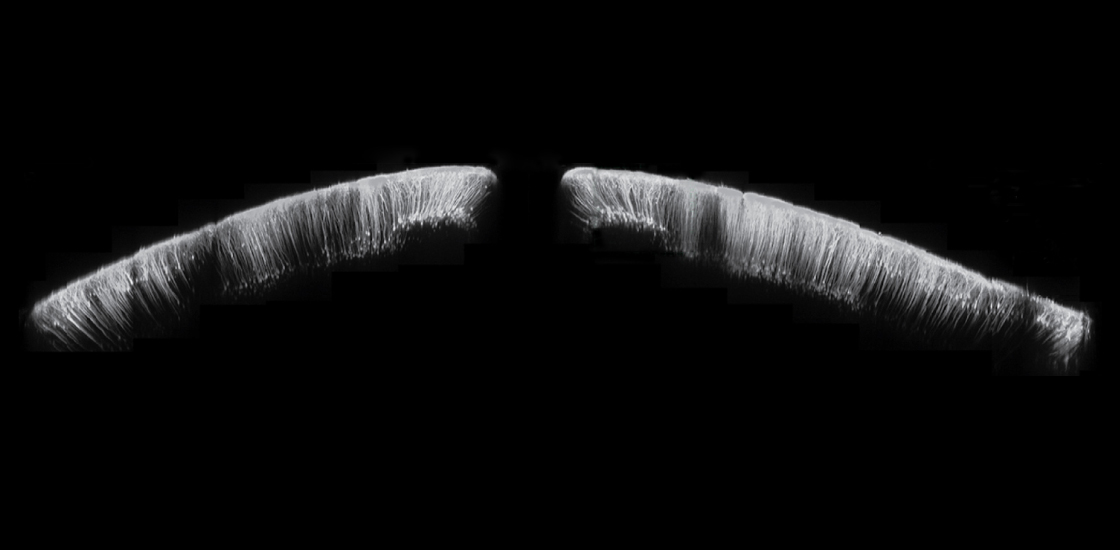
Clear view: Neurons in the outermost layer of a mouse brain are visible through a large glass window in the animal’s skull.
The new trapezoid-shaped window, described 20 December in Cell Reports, is gently curved to reduce stress on the brain. It is 8 millimeters long and 10 millimeters wide — big enough to view up to 40 mouse brain regions at once.
Researchers implanted the window in 18 mice. They scored the outline of the window into the skull and gently lifted one side so they could slip the glass window underneath. This procedure minimizes brain swelling.
The team then removed the piece of skull, fixed the window in place with glue and allowed the animal to recover. They examined the brains of four of the mice for signs of injury and inflammation, and saw none.
The researchers then used a microscope to visualize brain activity through the window. They could capture only a section of the panoramic view at a time, so they stitched several separate images together.
The resulting picture shows up to 50,000 neurons in the outer layers of the brain. Using a microscope that reveals deeper regions of the brain, the researchers could observe up to 1.1 million active neurons. The windows remain clear for at least 11 weeks.
By joining the discussion, you agree to our privacy policy.