THIS ARTICLE IS MORE THAN FIVE YEARS OLD
This article is more than five years old. Autism research — and science in general — is constantly evolving, so older articles may contain information or theories that have been reevaluated since their original publication date.
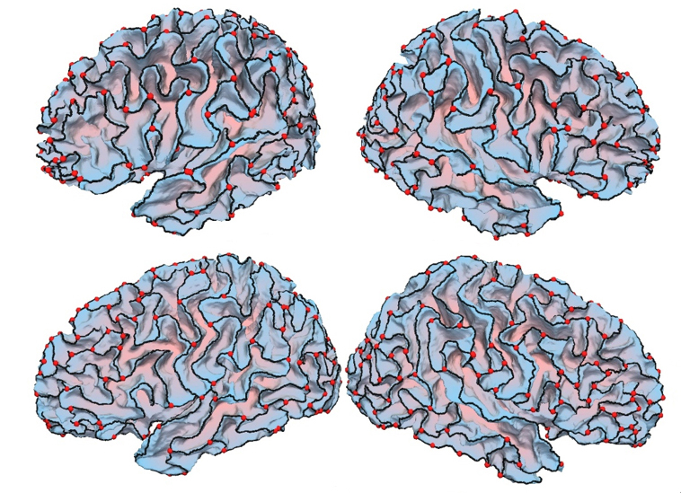
New software charts the crests of the folds that define the brain’s surface, or cerebral cortex, in people with autism1.
The cerebral cortex has distinctive crests and valleys, called gyri and sulci. These folds accommodate a large number of brain cells within the confines of the skull. Some studies suggest that people with autism have altered patterns of brain folding and unusually shaped sulci. But few studies have focused on gyri.
The new tool uses data from magnetic resonance imaging (MRI) scans to depict gyri in a mesh-like form, dubbed a gyral net. Two software packages identify the gyri in the scans. An algorithm then removes some of the detail, leaving a mesh made up of lines, each of which represents a ridge.
The program reveals characteristics of the mesh, such as the length of the lines, how straight they are and the number of times they intersect with other lines. It also approximates the length of neuronal connections. Creating a gyral net from MRI scans takes about 15 minutes for each hemisphere.
The researchers tested their tool on 40 MRI scans each from three people. The gyral nets for each person were similar to one another, showing that the software produces consistent results.
They then used the tool to compare the gyral nets of 185 people with autism with those of 252 controls. The scans came from a data-sharing resource called the Autism Brain Imaging Data Exchange.
Overall, the crests in people with autism are more convoluted than those in controls, the analysis found. And the paths between two points within a region were shorter, on average, in the autism brains, suggesting that these brains are tightly connected at a local level. Both observations are in line with previous research.
The researchers plan to tweak the algorithms to help the tool process brain scans faster. Scientists could use gyral nets to see whether certain patterns of brain folding track with autism behaviors.
By joining the discussion, you agree to our privacy policy.