THIS ARTICLE IS MORE THAN FIVE YEARS OLD
This article is more than five years old. Autism research — and science in general — is constantly evolving, so older articles may contain information or theories that have been reevaluated since their original publication date.
A new method allows researchers to rapidly map the routes of thousands of neurons in the brain1. The technique, described 7 September in Neuron, could reveal the arrangement of individual neurons in networks that underlie behaviors altered in autism.
Neurons send information through long projections called axons. These projections can be short, connecting neighboring cells, or long, linking distant brain regions.
Researchers can visualize axons by infecting neurons with a virus that emits a colorful glow under a microscope. But visually tracing each cell’s projection through large swaths of brain tissue takes months, and having a limited palette of fluorescent proteins means researchers can track only 5 to 10 cells in a single experiment.
With the new tool, called MAPseq, researchers can quickly — in less than a week — deduce the paths of thousands of neurons by swapping color codes for genetic barcodes. Viruses deliver the barcodes, which are random RNA sequences, into neurons.
The viruses also carry the code for a protein designed to transport RNA molecules to the end of an axon. When the virus infects a neuron, the cell produces the protein, which shuttles the RNA down the axon.
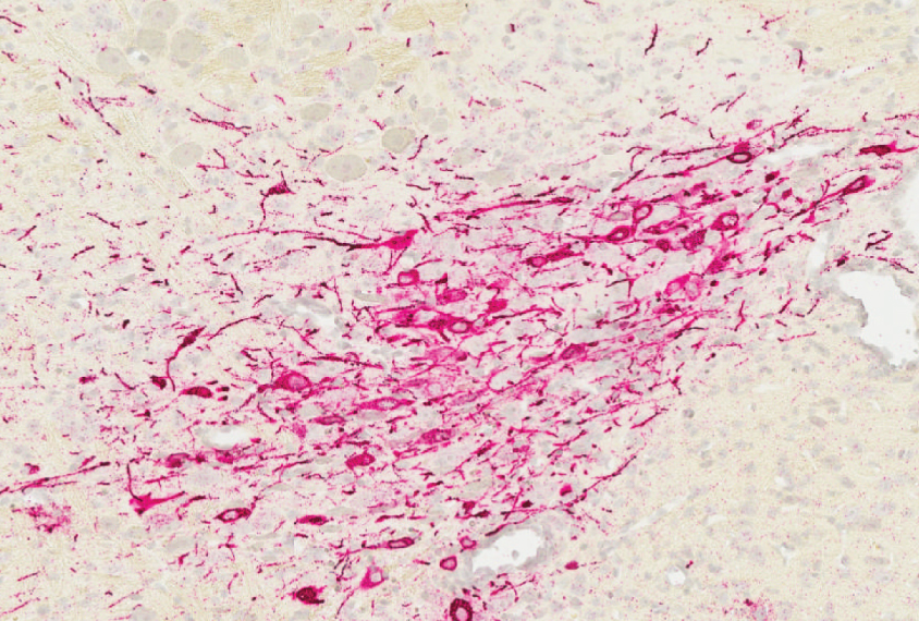
In addition, the viruses express a green fluorescent protein, allowing the researchers to see which cells have picked up a barcode.
Building bridges:
Researchers injected the viruses into the brainstem of living mice. Each virus infects one cell, so that each neuron receives a unique barcode.
Two days later, the researchers dissected the mouse brains. Under a microscope, 995 neurons glowed green, suggesting they had picked up a barcode.
The researchers then dissected the brain into five distinct brain regions and divided these regions into ultra-thin slices. They extracted RNA from each slice and performed automated high-throughput sequencing to determine which barcodes were present. They plotted the progression of individual barcodes through the slices to see where each cell’s axon ended up.
Their findings reveal that neurons in the locus coeruleus area of the brainstem, which is active in the stress response, extend to parts of the cerebral cortex, the brain’s outer layer. These neurons also connect to the olfactory bulb, a region involved in smell. The researchers did not find axons bridging the brainstem and the striatum, which governs motivation and movement.
Researchers can generate billions of unique barcodes, enough to tag every neuron in the human brain. Future versions of the tool may allow scientists to determine whether particular neuronal junctions boost or dampen brain activity.
By joining the discussion, you agree to our privacy policy.