THIS ARTICLE IS MORE THAN FIVE YEARS OLD
This article is more than five years old. Autism research — and science in general — is constantly evolving, so older articles may contain information or theories that have been reevaluated since their original publication date.
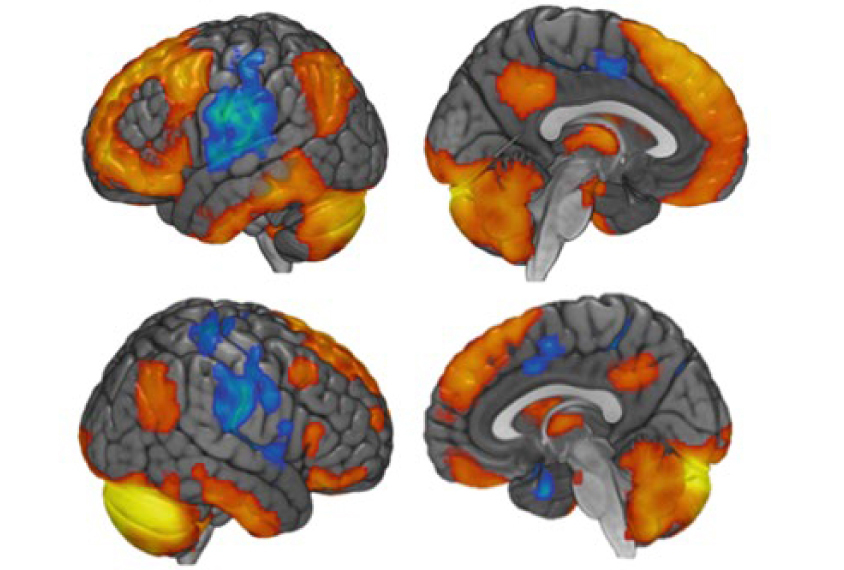
New evidence from both people and mice points to a part of the cerebellum that processes social information as being critical in autism1.
The findings indicate that this region, called the right crus I (RCrusI), is disrupted in children with autism. In mice, suppressing activity in the region can cause social problems and repetitive behaviors reminiscent of the condition; stimulating the region reverses the social problems.
The researchers were also able to artificially stimulate the area in typical adults — hinting at a therapy for autism.
Various studies have pointed to a role for the cerebellum in autism. For example, people with autism have fewer Purkinje neurons, which inhibit brain activity, in parts of the cerebellum2. The new work narrows the area of interest to the RCrusI.
“These findings are incredibly exciting and provide strong evidence that this region is an important player,” says Catherine Stoodley, associate professor of psychology at American University in Washington, D.C., who co-led the study.
The cerebellum governs motor coordination. It also plays a role in language, emotion regulation and other cognitive processes. Although the specific function of RCrusI isn’t known, earlier work suggested that it is involved in social and cognitive processing. The new findings also indicate that the RCrusI plays a part in social interactions.
The work also supports the use of mice for studying mechanisms involved in autism.
“You could have argued that the underpinning of social function in mice and rats is really not the same as in humans; this work kind of flies in the face of that,” says Emanuel DiCicco-Bloom, professor of neuroscience and cell biology at Rutgers University in New Jersey, who was not involved in the study.
Circuit control:
People with autism have less gray matter — tissue composed of neuronal cell bodies — in the RCrusI than controls do, according to a 2015 study3. But studies have not suggested a causative role for the RCrusI in autism until now, says Peter Tsai, assistant professor of neurology at the University of Texas Southwestern in Dallas, who co-led the study.
The team looked at the region in 34 typical adults. Using functional magnetic resonance imaging (fMRI), they looked for brain areas that become active in tandem with the RCrusI.
The RCrusI is functionally connected to some regions associated with autism, the researchers found. For example, it connects to the frontoparietal network, which is involved in high-level thought processes, such as attention4.
As proof of principle, the team then showed that they could stimulate brain activity in the RCrusI using transcranial direct current stimulation — which delivers an electrical current to the brain — for about 20 minutes in 19 typical adults. (The other adults received a sham treatment.)
The team then used fMRI to examine brain activity in the RCrusI in 81 children with autism, aged 8 to 13 years. They also looked at activity in a region of the frontoparietal network called the inferior parietal lobule (IPL).
These children showed greater communication between the RCrusI and the left IPL than did 81 typical children, the researchers found. The study appeared 30 October in Nature Neuroscience.
Connection cues:
In parallel, the team also examined structural connections in the brains of mice missing a gene called TSC1 in their Purkinje neurons. TSC1 is mutated in people with tuberous sclerosis, a condition often accompanied by autism. Mice missing TSC1 in their Purkinje neurons show repetitive behaviors, such as excessive grooming.
The TSC1 mice have atypical connections between the RCrusI and several other areas, including the frontoparietal network, the researchers found.
The researchers then disrupted Purkinje neurons in the RCrusI in typical mice. They injected the mice with a virus that targets these inhibitory cells. The virus carries a protein receptor that, when activated by a drug, dampens the cells’ activity.
Mice injected with the virus become less social: They do not show a preference for approaching another mouse over an object, as controls do. They also begin grooming themselves excessively, and cannot switch strategies when trying to navigate a maze, indicating that they have trouble with mental flexibility.
The researchers then injected a different receptor that enables them to stimulate Purkinje neurons in the mice’s RCrusI. This intervention restores the mutants’ interest in other mice but has no effect on their grooming or mental flexibility.
Stimulating the RCrusI in people might similarly improve social skills in those with autism, Stoodley says. “It gives us a potential therapeutic target down the road.”
However, using the same approach in the left crus I region does not produce these changes. That specificity is surprising, says Samuel Wang, professor of molecular biology at Princeton University, who was not involved in the study.
“Mice are not known to have consistent hemispheric dominance, which is thought to be connected with handedness in humans,” Wang says. “So we will have to see if that result is replicated.”
Stoodley and her team plan to use the stimulation technique on 15 adults with autism to probe the role of the cerebellum — and of the RCrusI — in the condition.
By joining the discussion, you agree to our privacy policy.