THIS ARTICLE IS MORE THAN FIVE YEARS OLD
This article is more than five years old. Autism research — and science in general — is constantly evolving, so older articles may contain information or theories that have been reevaluated since their original publication date.
By matching gene expression patterns in postmortem brains to imaging data from living individuals, researchers have uncovered 38 genes that may collaborate to control neural activity in the brain.1 Nine of the genes are known autism candidates.
The findings, published 18 November in Neuron, could help uncover the molecular underpinnings of brain activity, says lead investigator Genevieve Konopka, assistant professor of neuroscience at University of Texas Southwestern Medical Center in Dallas. The results may also help researchers understand how these genes might contribute to autism.
“If we can identify specific genes that are important for brain activity in autism or other disorders, these could be potential targets for drug therapies way down the line,” Konopka says.
Konopka and her colleagues merged data from two disparate sources: brain scans from 282 healthy people, performed while their brains were at rest — meaning not actively engaged in any task — and postmortem brain tissue from 50 adults with no known genetic disorders.
This blending of genetic and imaging data is an innovative approach to understanding the causes of complex brain disorders, says Paul Thompson, professor of neuroimaging at the University of Southern California.
“You can certainly go through the genetic sequence and look for culprits that affect our vulnerability to these diseases, but you never quite know the mechanisms of the genes promoting disease,” says Thompson, who organizes an international repository of imaging and genomics data called the ENIGMA Consortium. “In this case, brain scans are the missing key.”
Dynamic connections:
Previous studies have searched for altered gene expression in postmortem brain tissue from people with autism. But outside the context of a living, functioning brain, it’s difficult to know if these abnormalities are meaningful.
In the new study, the researchers tried to address this limitation.
“The people being imaged are alive, and the gene expression data are from postmortem brains. But by using large enough sample sizes, we hoped to approach a population-level understanding and find correlation between these two,” Konopka says. “And indeed that’s what we found.”
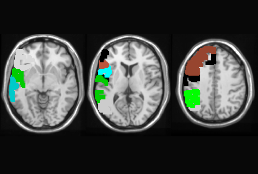
Konopka and her team combed through functional magnetic resonance imaging (fMRI) data from two public datasets. They examined patterns of brain activity in six regions of the cerebral cortex, the brain’s outer layer, in images taken while the individuals rested in the scanner. These regions include part of the prefrontal cortex and five others known to be involved the brain’s ‘resting state.’ The researchers reasoned that this state of the living brain is most similar to that of the brain after death, allowing a valid comparison between the groups.
Genomic signature:
They then sequenced RNA from the same regions of the cerebral cortex in postmortem brains from adults with no known genetic disorders, ranging in age from 33 to 49. The scientists increased their sample size by combining their results with published data from the same regions in the BrainSpan Atlas, a repository of gene expression profiles in postmortem brains.
A side-by-side comparison of fMRI and gene expression patterns revealed 38 genes whose expression levels track with brain activity in the six regions. The genes appear to act in concert, Konopka says. Of the 38 genes, 12 increase in expression, and the other 26 decrease, as brain activity surges.
“The exciting thing is that there really is a genomic signature to brain activity, and it seems to be driven by neurons,” Konopka says. She notes that 4 of the 38 genes are markers of interneurons, cells that act as connectors between other neurons, sometimes across brain regions.
In neurons, most of the genes associated with brain activity were expressed at levels at least twice as high as in other cell types, such as endothelial or glial cells, which line blood vessels and support neurons, respectively.
Autism link:
A 2014 analysis of postmortem brain tissue from a different team uncovered nine genes from scattered parts of the cerebral cortex that are expressed at lower levels in people with autism than in the general population. The list of 38 genes in the new study includes these genes. The list also includes SCN1B, a sodium channel regulator implicated in the seizure disorders epilepsy and Dravet syndrome, both of which are associated with autism.
Many of the genes are consistently expressed across all areas of the cerebral cortex in an adult brain, according to a report in December in Nature Neuroscience2. This suggests the genes work to maintain functional connections between various regions of the brain.
“This affirms the importance of these genes in establishing functional connectivity,” says Michael Hawrylycz, investigator at the Allen Institute for Brain Science in Seattle, who led the Nature paper.
A study published this summer in Science involved a similar combination of fMRI in healthy individuals and genetic analyses of brain tissue but encompassed 88 brain regions, including 3 regions included in the new study3. The Science study identified SCN1B and three other genes on Konopka’s list, supporting the notion that dozens of genes operate behind the scenes to modulate the activity seen in brain scans. However, it did not finger any autism gene candidates.
“We’re all identifying a core set of genes even though we’re using different methods and different samples,” Konopka says. Her group plans to examine the genes in animal models to explore the relationship between gene expression and brain function, focusing particularly on autism candidates.
By joining the discussion, you agree to our privacy policy.