THIS ARTICLE IS MORE THAN FIVE YEARS OLD
This article is more than five years old. Autism research — and science in general — is constantly evolving, so older articles may contain information or theories that have been reevaluated since their original publication date.
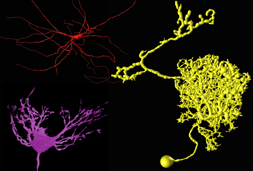
A computer program called NeuronStitcher can quickly and accurately sew a series of brain scans into a three-dimensional rendering. The open-source program, described today at the 2015 Society for Neuroscience annual meeting in Chicago, could help researchers trace the paths of individual neurons through the brain.
High-resolution microscopy can reveal the intricate details of neurons and their connections. But in order to visualize a neuron from its cell body to the tip of its axon, researchers have to collect a series of images at different tissue depths. They then have to stitch these snapshots together like a quilt.
NeuronStitcher automates this tedious and time-consuming process.
“We have more and more data coming in from high-resolution imaging techniques,” says Hanbo Chen, who developed the tool as a visiting student in Hanchuan Peng’s lab at the Allen Institute for Brain Science in Seattle. “It’s a tool to process the increasing amounts of imaging data,” he says.
NeuronStitcher works by identifying tiny spots on successive images called ‘border tips.’ These spots represent fragments of neurons that are either exiting or entering the focal plane. By matching border tips in serial sections of tissue, NeuronStitcher can follow a neuron’s tangled path.
“This is very useful for simulating neural networks,” says Chen.
Chen and his colleagues tested the tool by scanning a piece of mouse brain with confocal microscopy and then cutting it into three sections. They scanned each of the sections separately and stitched the snapshots together with the software. The program reconstructed the intact tissue with more than 95 percent accuracy.
The researchers also used NeuronStitcher to process data from 10 pairs of tissue sections taken from different experiments. The program correctly matched more than 80 percent of the border tips in this hodgepodge of images.
Although the program is automated, it gives researchers the opportunity to evaluate and correct the results manually. The program is freely available as a plug-in from the Allen Institute’s BigNeuron project.
For more reports from the 2015 Society for Neuroscience annual meeting, please click here.
By joining the discussion, you agree to our privacy policy.