THIS ARTICLE IS MORE THAN FIVE YEARS OLD
This article is more than five years old. Autism research — and science in general — is constantly evolving, so older articles may contain information or theories that have been reevaluated since their original publication date.
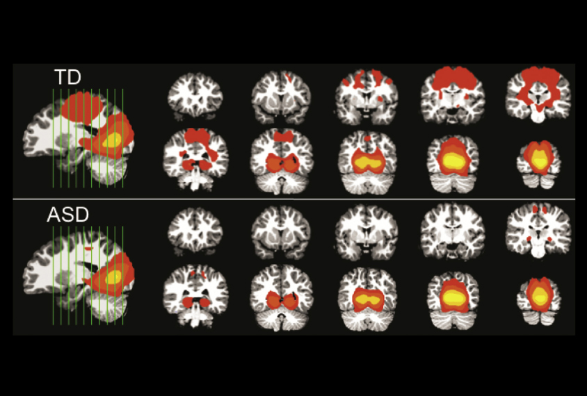
Scanning young children with autism as they sleep reveals reduced synchrony between a brain region that processes emotions and structures involved in social communication1.
The findings, published in the September issue of the Journal of the American Academy of Child and Adolescent Psychiatry, add to a growing body of evidence that connections between brain areas are altered in autism.
Researchers measure connectivity by identifying brain areas that activate together as a person rests in a magnetic resonance imaging (MRI) scanner. This synchronization indicates the areas are functionally linked.
Studies suggest that the brains of school-age children with autism are more strongly connected than are those of controls. But by then, any autism treatments a child has received may have already influenced his brain activity, says lead investigator Christine Nordahl, assistant adjunct professor of psychiatry and behavioral sciences at the University of California, Davis.
“You don’t know if you’re looking at the effect of intervention or the effect of life as opposed to the neural basis of autism,” Nordahl says.
To sidestep this problem, Nordahl and her colleagues scanned a group of children with autism, most of whom were aged 2 to 4 years. Children this young are notoriously fidgety, and any amount of movement can skew the images. So the researchers scheduled the scans for bedtime and allowed the children to fall asleep at the clinic. They then slipped the children into the MRI machine and scanned them as they slumbered.
This lights-out approach is not directly comparable to scanning participants who are inactive but awake. But it gives researchers an unprecedented glimpse of brain connectivity during a stage of development that has been difficult to study, says Nouchine Hadjikhani, associate professor of radiology at Harvard University, who was not involved in the work.
“I can’t imagine the time and effort that went into that study,” she says. “That’s really remarkable.”
Bedtime stories:
The researchers scanned the brains of 43 boys with autism and 29 controls. They found that children with autism show less synchrony between the brain’s emotion hub, called the amygdala, and regions that play a role in social communication and repetitive behaviors, such as the medial prefrontal cortex and striatum, respectively.
Children with the lowest levels of synchrony have the most severe autism features as measured by the Autism Diagnostic Observation Schedule, a widely used test for the condition.
“It’s always nice when you can relate brain findings with behavior, which happens more rarely than you would think,” Nordahl says.
To see if the findings are autism-specific, the researchers gauged how well a visual brain region that is not implicated in autism synchronizes with other areas. They unexpectedly found that children with autism also show less connectivity than controls do between the visual cortex and brain regions involved in movement and touch.
The decrease in connectivity tracks with an increase in sensory sensitivity, according to a questionnaire called the Short Sensory Profile. Enhanced sensory sensitivity often accompanies autism.
However, the researchers did not look at synchronization between other brain regions. Finding brain regions that do not show altered connectivity in children with autism would help to confirm that the new results are specific to the condition, says Hadjikhani.
Nordahl and her team plan to scan the same group of participants over time to see how connectivity changes with age. This work may reconcile the findings in preschoolers with those in older children. The team is tracking some of the first children to join the study, who are now as old as 12.
A long-term study of brain connectivity in autism would be “brilliant,” Hadjikhani says. “They are sitting on a gold mine if they have all these kids that can tolerate being in a scanner.”
By joining the discussion, you agree to our privacy policy.