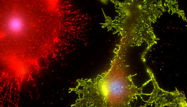
Riccardo Cassiani-Ingoni / Science Source
THIS ARTICLE IS MORE THAN FIVE YEARS OLD
This article is more than five years old. Autism research — and science in general — is constantly evolving, so older articles may contain information or theories that have been reevaluated since their original publication date.
Editor’s Note
This article was originally published 18 October 2015, based on preliminary data presented at the 2015 Society for Neuroscience annual meeting in Chicago. We have updated the article following publication of the study 10 October 2018 in Neuron1. Updates appear below in brackets.
A protective molecular tag on neurons can prevent microglia, the brain’s immune cells, from trimming away their connections with other neurons. The unpublished findings, presented Sunday at the 2015 Society for Neuroscience annual meeting in Chicago, reveal how microglia may target or ignore certain neuronal connections for pruning.
During development, the brain starts out with more neuronal connections, or synapses, than it needs. Shortly after birth, weak or unused connections are disposed of, and the remaining ones are strengthened. This pruning process is crucial for proper brain wiring and function, and is thought to go awry in autism.
Microglia, which engulf pathogens, dead cells and other debris in the brain, contribute to synapse pruning during development. A 2012 study showed that these cellular scavengers rely on a certain molecular cue to home in on and gobble up synapses that are weak and unused. Mice that lack this ‘eat me’ signal, or its receptor, are deficient in pruning and have too many neuronal connections.
But it has remained unclear whether microglia rely on additional molecular tags to keep their feasting in check. “Maybe they need cues to prevent them from engulfing the synapses that need to be maintained,” says Emily Lehrman, a postdoctoral fellow in Beth Stevens’ lab at Children’s Hospital Boston, who presented the findings.
Pruning protector:
The researchers focused on a molecule [called CD47] that protects healthy cells from being devoured by the body’s immune system, reasoning that this same molecule might serve a similar function in the brain.
They found that the protective tag is highly expressed at synapses in the visual system of mice at 5 days of age, when synapse pruning peaks in this brain region. During this same time period, expression levels of the receptor for this tag peak in microglia.
Deleting the protective tag from mice increases microglia’s ‘appetite’ for synapses during brain development and decreases the number of neuronal connections the mice have at 2 months of age. Deleting the receptor for the protective tag has similar effects.
The findings suggest that the protective tag and its receptor work together to regulate synapse pruning by microglia during development. One open question is whether this process is regulated by the activity of the target neurons.
“We know from many studies that activity is important for refinement, and that it’s generally the strong synapses that are maintained, and the weak synapses are removed,” Lehrman says. The researchers are investigating whether the protective tag preferentially marks strong connections.
“The data is indeed very interesting,” says Jonathan Kipnis, director of the Center for Brain Immunology and Glia at the University of Virginia in Charlottesville. Kipnis was not involved in the study but attended the presentation. “There possibly are many other molecules with similar or overlapping functions.”
[When the researchers suppressed neuronal activity in one eye in mice, they found that CD47 levels decreased in the signal-sending end of neurons of that eye and increased in those of the more active eye. They also found that microglia preferentially consume the synapses in the less active eye.
The researchers then tested microglia’s preference for active synapses in mice lacking CD47. They found that microglia could no longer distinguish between the normal eye and the one with suppressed neuronal activity. The findings suggest that CD47 levels play a role in microglia’s discriminating palate.]
For more reports from the 2015 Society for Neuroscience annual meeting, please click here.
By joining the discussion, you agree to our privacy policy.