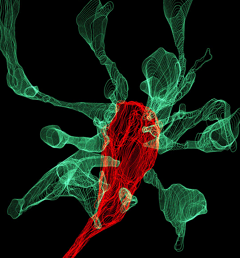
L. Weinhard, EMBL Rome
THIS ARTICLE IS MORE THAN FIVE YEARS OLD
This article is more than five years old. Autism research — and science in general — is constantly evolving, so older articles may contain information or theories that have been reevaluated since their original publication date.
Editor’s Note
This article was originally published 14 November 2016, based on preliminary data presented at the 2016 Society for Neuroscience annual meeting in San Diego. We have updated the article following publication of the study 26 March 2018 in Nature Communications1. Updates appear below in brackets.
Microglia come into frequent contact with synapses, the connections between neurons, but they appear to nibble on them rather than engulf and digest them.
The unpublished findings, presented today at the 2016 Society for Neuroscience annual meeting in San Diego, challenge the popular idea that microglia shape the brain by eliminating synapses.
“There are many groups that are trying to see this elimination happening,” says Laetitia Weinhard, a graduate student in Cornelius Gross’s lab at the European Molecular Biology Laboratory in Monterotondo, Italy, who presented the findings. “I think if nothing worked out so far, it’s because it’s really not easy to see or it’s not happening the way we thought.”
Microglia are known to be in close contact with neurons in the developing brain. Some researchers have found synaptic proteins inside microglia during periods of intense synapse pruning in the brain, suggesting that microglia trim synapses.
Weinhard and her colleagues used microscopy to look at live cells in brain tissue. Their goal was to see microglia in the act of eating synapses.
The researchers obtained slices of the hippocampus from the brains of 3-day-old mice. The hippocampus is a brain region involved in learning and memory that undergoes tremendous changes in synapse density throughout life. The researchers bred the mice so that their microglia glow red and some of their neurons glow green.
The researchers grew the brain slices in culture for 10 days to give the cells time to adjust to new conditions. They then scanned the samples using light-sheet microscopy, a technique that collects data from a large plane of tissue. They captured one picture every minute for three hours and assembled the images into videos.
During the imaging sessions, the microglia extended just as many projections as they retracted, a sign that the researchers’ microscopy technique does not interfere with the cells’ activity. “If the conditions are not proper, microglia tend to retract more than to extend the [projections],” says Weinhard.
Little bites:
The researchers then turned their attention to the interactions between microglia and dendritic spines, tiny neuronal branches that receive signals from other neurons. They focused on [31] spines that the microglia had come into contact with during the three-hour time frame of the videos. Microglia had touched most of those spines more than once.
[About 10 percent] of the spines vanish partway through the videos, but their disappearance never coincides with the presence of a microglial cell, the researchers found. Interestingly, about 50 percent of the spines appear to become stretched as microglia pull away after touching them. The microglia seem to remove tiny pieces of the spine, but the structure looks otherwise intact. Microglia may “take nibbles and bits off the head of the spine,” Weinhard says.
To get a closer look at these interactions, the researchers isolated chunks of hippocampus from the mouse brain and preserved them to freeze all of the cells in place. They then looked at more than 8,000 individual spines using confocal microscopy.
About [1] percent of the spines appear to be surrounded by a microglial cell, but all of these spines remain physically attached to a neuron.
Taken together, the findings cast doubt on the idea that microglia eliminate synapses by devouring them, Weinhard says.
Other scientists are more sanguine, however: The researchers focused on the signal-receiving ends of synapses, but microglia preferentially eat the signal-sending ends, says Beth Stevens, assistant professor of neurology at Harvard University.
Stevens’ team has looked at microglia in the visual system, whereas the new study focused on the hippocampus, which is less well understood, Stevens says. “It is possible that they are missing the correct time point and location of developmental spine pruning in the hippocampus, as this has not been as well established as in sensory systems,” Stevens says.
To confirm her theory that microglia are only nibbling small pieces of synapses, Weinhard says she is trying to observe these interactions at even higher resolution using electron microscopy.
[Weinhard and her colleagues re-examined the data to focus on the signal-sending ends of synapses. They found that microglia take small bites out of the signal-sending ends but do not consume the entire structure.
None of this nibbling activity was altered in brain slices from mice lacking a receptor called CR3, which some reports indicate microglia use to trim away certain synapses. This finding contrasts with studies exploring the role of microglia in trimming synapses in the visual system.
In these follow-up experiments, the researchers did not find parts of the signal-receiving ends inside microglia, suggesting microglia do not nibble these ends after all. Instead, they saw that this part of the synapse extends a thin protrusion when microglia contact it. Of 21 spines that extended protrusions after microglia contact, 5 later changed their synaptic partner — a phenomenon called ‘spine switching.’ This process is thought to be important for circuit remodeling, which underlies learning and memory.]
For more reports from the 2016 Society for Neuroscience annual meeting, please click here.
By joining the discussion, you agree to our privacy policy.