THIS ARTICLE IS MORE THAN FIVE YEARS OLD
This article is more than five years old. Autism research — and science in general — is constantly evolving, so older articles may contain information or theories that have been reevaluated since their original publication date.
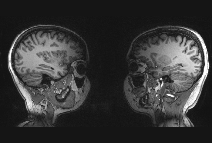
A functional magnetic resonance imaging (fMRI) scanner that is designed to simultaneously measure brain activity in two people holds promise for capturing the nuances of social interaction and nonverbal communication.
The machine, described yesterday at the 2015 Society for Neuroscience annual meeting in Chicago, is just one of several efforts in the past few years to assess the brain during real-world social interactions — a key to understanding the social difficulties of people with autism.
An earlier two-person MRI method used a pair of magnetic coils to image two brains, whereas the new machine has two arrays of smaller coils. The array design is more sensitive to activity in the cerebral cortex, the brain’s outer rind. This may make the new machine better able to capture cortical functions such as social communication and cognition, says Ville Renvall, a postdoctoral researcher in Lauri Nummenmaa’s lab at Aalto University in Finland, who presented the work.
Renvall and his colleagues had to figure out how to fit two heads and two arrays inside one scanner. But in the end, “We actually get pretty nice, full two-brain coverage from these images,” Renvall says.
The researchers tested their apparatus with 10 pairs of young adults who lay down facing each other in the bore of the scanner — a setup Renvall describes, understatedly, as “cozy.”
They scanned the pairs’ brains while each member of a pair took turns lightly tapping an index finger on the other person’s lower lip. During this process, the person doing the tapping shows activation in motor areas of the cortex, while the person receiving the touch shows activation in sensory areas.
“Mostly the responses flip from one brain to the other,” says Renvall. “The role that they are taking is definitely making a difference.”
When the participants hear an instruction through the scanner’s intercom system to start or stop tapping, both members of the pair show activation in parts of the cortex that process sound.
Intimate setting:
The results were as expected, showing that the new scanner accurately picks up brain activity during different phases of a social task. “I’m pretty happy that this dual-coil fMRI seems to be already usable for studying social interaction,” Renvall says.
But applying this or similar methods to individuals with autism could pose challenges. Renvall’s setup is undeniably intimate: The participants rest with their faces mere centimeters apart. People with autism might find being in such close proximity hard to handle, as face-to-face communication can be overwhelming to them even at a normal distance.
Renvall says there may be ways to lessen potential anxiety, however. “I think we can set it up so that they can be made more comfortable with it,” he says. For example, people could be allowed to familiarize themselves with the equipment beforehand. Emphasizing the experimental, laboratory nature of the setting could also take some social pressure off.
But before inviting individuals with conditions such as social anxiety or autism into the scanner, Renvall says he needs to make some technical tweaks. He says he would like to apply better ways of capturing and processing images to the two-person setting, and improve the synchronization of measurements of the two brains.
For more reports from the 2015 Society for Neuroscience annual meeting, please click here.
By joining the discussion, you agree to our privacy policy.