THIS ARTICLE IS MORE THAN FIVE YEARS OLD
This article is more than five years old. Autism research — and science in general — is constantly evolving, so older articles may contain information or theories that have been reevaluated since their original publication date.
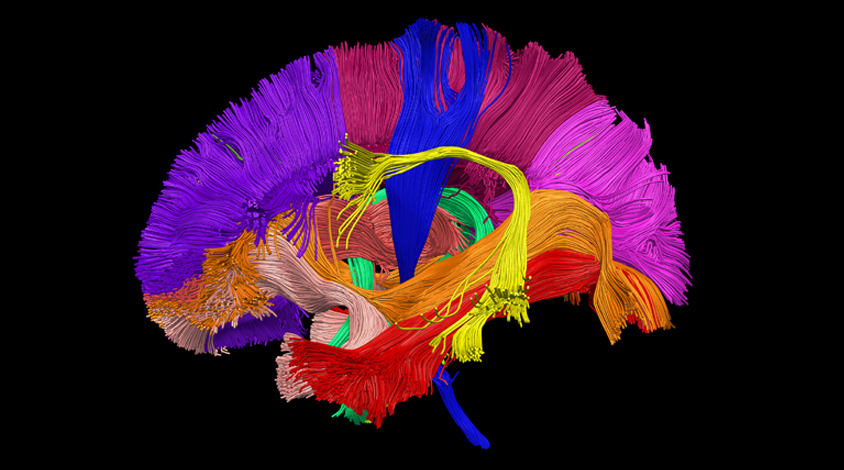
Bundles of nerve fibers that bridge brain areas develop rapidly during the first six months of life. Bundles that connect language regions mature more slowly than those linking motor regions.
The unpublished results were presented yesterday at the 2017 International Meeting for Autism Research in San Francisco, California. Researchers could use the results as a benchmark to investigate how brain development is altered in autism.
“We hope that by doing the same analysis in children with [autism] we can identify tracts that may be altered or derailed early in development,” says Longchuan Li, co-director of the Pediatric Neuroimaging Core at Marcus Autism Center in Atlanta, Georgia.
Li and his colleagues used diffusion tensor imaging (DTI) to visualize 11 different nerve bundles, called white matter tracts, in 33 typically developing infants, 10 of them girls.
DTI is based on the diffusion of water across the nerve tracts, which indicates their integrity. Tracts typically become more intact, and robust, with age, but some studies suggest children with autism are born with atypically mature tracts that undergo a kind of structural regression over time.
The analysis revealed that all 11 of the white matter tracts in a typical infant’s brain become more mature over time, but certain tracts mature faster than others. For instance, the pyramidal tract, which connects the motor cortex to the spinal cord, matures more quickly than the arcuate fasciculus, which connects regions of the brain’s surface involved in language. This makes sense, Li says, because children typically reach motor milestones before language ones.
Sleepy scans:
Each infant underwent up to three scans between birth and 6 months of age. The youngest baby scanned in the study was 20 days old.
The scans were eight minutes, on average, and took place while the babies were sleeping. Their mothers nursed or rocked them to sleep in a dark room with the scanner, and then placed them on a platform that moved them into the scanner. The babies wore headphones to block out the noise of the scanner.
The researchers used an integrity measure called fractional anisotropy, which reflects the water diffusion along and between nerve fibers. Their analysis of this measure revealed that all of the brain’s nerve tracts develop most rapidly during the first three months of life. Development slows thereafter. The rate of change to the integrity of the tracts in the brain at 1 month is six times higher than the rate of change at 6 months.
Li suspects that 3 of the 11 tracts are altered in autism: the pyramidal tract, the arcuate fasciculus and the uncinated fasciculus, which runs through the brain’s emotion-processing center, the amygdala. These tracts underlie behaviors that are altered in autism, such as motor, language and social ability.
For more reports from the 2017 International Meeting for Autism Research, please click here.
By joining the discussion, you agree to our privacy policy.