THIS ARTICLE IS MORE THAN FIVE YEARS OLD
This article is more than five years old. Autism research — and science in general — is constantly evolving, so older articles may contain information or theories that have been reevaluated since their original publication date.
A technique that expands brain tissue to four times its original size lets researchers spot and sequence individual messenger RNAs (mRNAs) — the genetic blueprints for protein production. The method, described today at the 2015 Society for Neuroscience annual meeting in Chicago, could reveal how a single neuron makes thousands of connections, called synapses, with different roles in the brain.
“In order to be different, [synapses] need to have different proteins,” says Shahar Alon, a postdoctoral fellow in Edward Boyden’s lab at the Massachusetts Institute of Technology in Cambridge. “But we don’t know if this [difference] is also at the level of the mRNA.”
Earlier this year, Boyden’s lab reported a technique called expansion microscopy. The method uses a water-absorbing gel found in disposable diapers to stretch brain tissue while preserving its cellular structures1.
“You can get super resolution just by using an ordinary microscope,” Alon says.
In the new study, Alon and his colleagues combined expansion microscopy with fluorescent in situ sequencing (FISSEQ), a technique developed in George Church’s lab at Harvard Medical School2. The method works by binding a series of colorfully labeled bases to the mRNA. By recording the resulting sequence of colors, researchers can deduce the mRNA sequence.
The researchers call the combined technique ‘expansion sequencing,’ or ExSEQ.
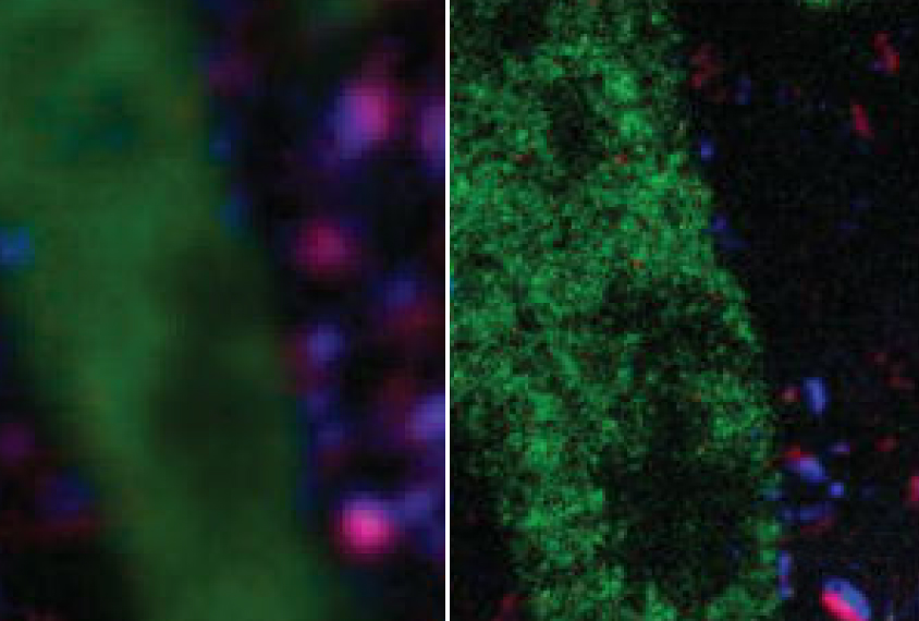
Mapping messages:
To put ExSEQ to the test, Alon and his colleagues used a fluorescent marker to label mRNA in a slice of mouse brain. They then expanded the tissue so they could distinguish between individual mRNAs.
After pinpointing the mRNA, which glowed green in the tissue, the researchers used FISSEQ to sequence it. They were able to detect mRNAs involved in a range of cellular processes, such as forming synapses and guiding neuronal projections to their targets.
Although the technique doesn’t capture every mRNA, it appears to catch a representative sample. Alon says it would be especially useful for researchers interested in a specific cellular structure, such as the synapse.
“If you have only a few copies of messenger RNA, but they are located in space in a very precise location, like the synapse, I think this method can be good,” he says.
Alon plans to use ExSEQ to measure mRNA expression in different synapses on the same cell.
“We know that every neuron can have thousands of synapses, and we know that these synapses can be functionally different,” he says. “But we don’t know what is the exact mechanism that makes them different.”
For more reports from the 2015 Society for Neuroscience annual meeting, please click here.
By joining the discussion, you agree to our privacy policy.