THIS ARTICLE IS MORE THAN FIVE YEARS OLD
This article is more than five years old. Autism research — and science in general — is constantly evolving, so older articles may contain information or theories that have been reevaluated since their original publication date.
An imaging technique that lights up cells without involving fluorescent markers lets scientists see deep inside spheres of neurons called organoids.
Researchers used the technique to spot layers of immature neurons in organoids derived from the cells of people with Rett syndrome, a condition linked to autism. The unpublished results were presented yesterday at the 2016 Society for Neuroscience annual meeting in San Diego.
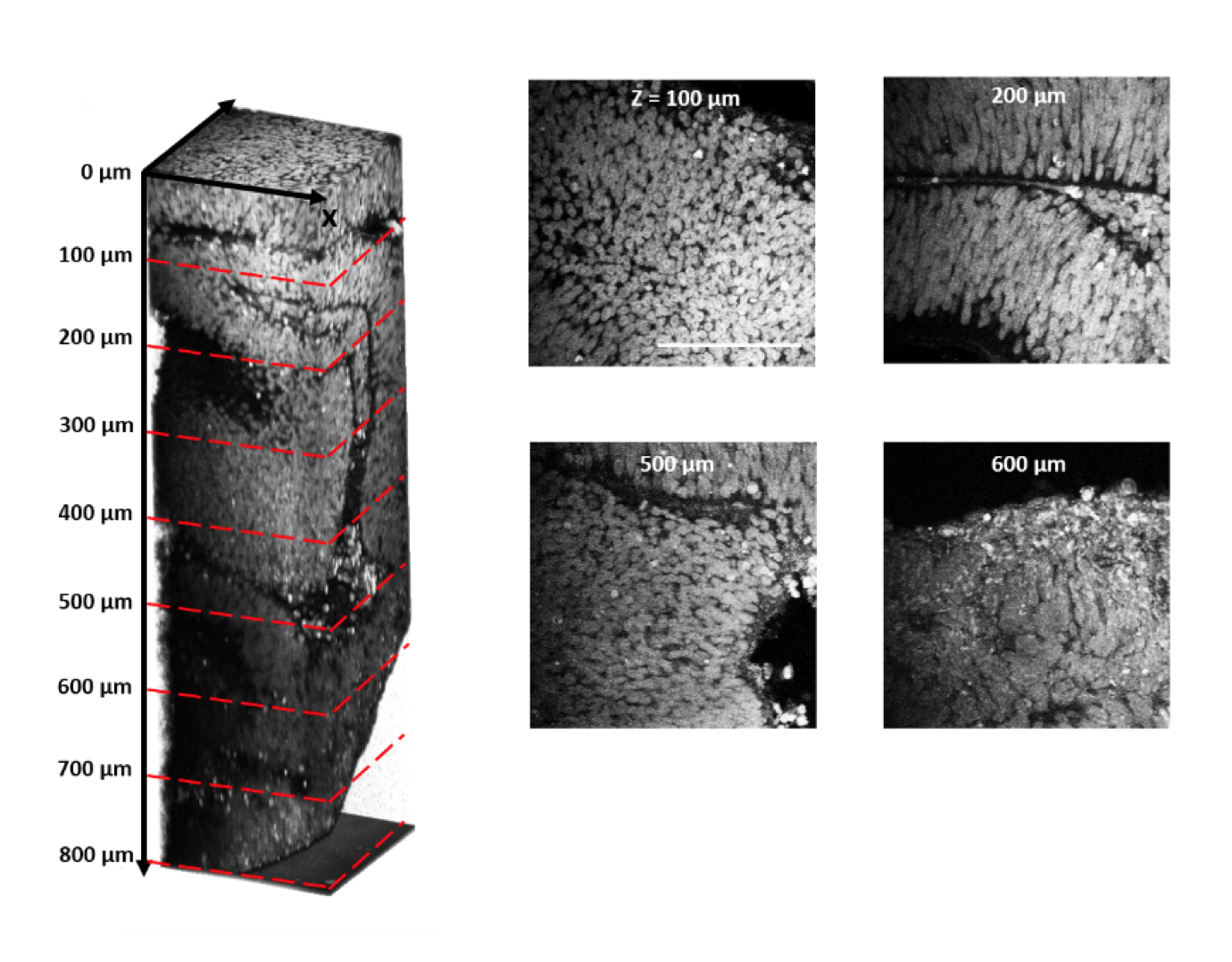
Traditional methods for imaging brain organoids, or ‘mini-brains,’ involve taking a snapshot of a thin cross-section of the tissue. But this fails to capture the organoids’ three-dimensional structure. And to see individual cells, researchers must label them with fluorescent markers. Delivering these labels to the center of a mini-brain and making them visible through the tissue layers is challenging.
Researchers have worked around the latter problem by rendering the tissue transparent. Even so, they cannot capture the glow of cells beyond a certain depth.
In the new method, researchers illuminate neurons throughout an organoid using a technique called third harmonic generation microscopy. It involves tuning a laser to light up cells with a defined set of physical properties, such as a particular shape, deep inside tissue.
“We are still in the early phases, but we think that we can use this to get a better idea of what’s happening inside [Rett organoids],” says Murat Yildirim, a postdoctoral researcher in Mriganka Sur’s lab at the Massachusetts Institute of Technology in Cambridge, Massachusetts. That understanding could provide clues to the causes of Rett syndrome, Yildirim says.
Label-free imaging:
Using traditional imaging techniques, Sur and his colleagues previously showed that Rett mini-brains have an unusually large number of structures that resemble fluid-filled brain cavities called ventricles. These structures look underdeveloped.
In the new work, the team first made the organoids clear. They then used a laser to shine a particular wavelength of light into the tissue. The light reflected off of the cells, revealing an area around the ventricles in the Rett organoids lined with what looks like immature progenitor cells that are still dividing. This area should consist of layers of mature neurons that have migrated outward.
“This corresponds to an immature cortex,” says Danielle Feldman, who worked on the project as a graduate student in the Sur lab. The Rett syndrome cells “are stunted in their differentiation,” Feldman says.
The researchers plan to use the new laser method to compare the structure of Rett organoids with that of organoids derived from typical individuals. Theoretically, they also could use the technique to visualize organoids as they develop.
For more reports from the 2016 Society for Neuroscience annual meeting, please click here.
By joining the discussion, you agree to our privacy policy.